Archive for the ‘Dogs’ Category

Mast Cell Tumours in dogs, what are they, and how can it be treated?

Mast cell tumours originate from Mast Cells

Mast cells are found in normal healthy dogs; they are white blood cells, found near blood vessels within the connective tissue. The cells are particularly equipped to catch germs and prevent them from spreading throughout the body, but they are also able to repair tissue and assist in creating new blood vessels. Essentially, they are pretty important as they play a key role in defending the body and keeping the immune system healthy and strong. They also release histamine and other compounds which are involved in allergic reactions. 
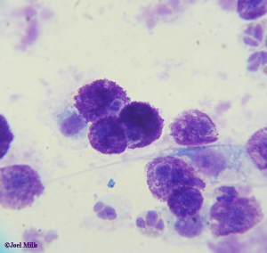
So, a mast cell tumour is…?

Mast cell tumours occur when cancerous changes occur in the mast cells, allowing them to multiply too quickly and out of control. Mast Cell Tumours are very variable in appearance,  from smooth, skin coloured swellings to angry looking masses with numerous, bumpy lumps. The lump may well appear inflamed and be itchy, causing your pet some irritation and discomfort. If the tumour is in a more progressive stage then vomiting and diarrhoea can occur due to the excess levels of histamine produced by the cells of the tumour.

Brachycephalic breeds (the ones with short snouts and wide heads e.g. Boxers, Pugs, Bulldogs etc.) are at a particularly high risk of developing these tumours. The perineum (the area between the genitals and anus) is a common site for mast cell tumours, but they can also occur on the limbs, face or anywhere on the body. If you notice a new lump anywhere on your pet, it is always worth getting it looked at by one of our vets.

Sometimes the tumour will grow quickly so it is important to contact us ASAP to discuss any new lumps appearing on your pet.  Histamine, released by Mast Cell Tumours, can cause swelling in the surrounding skin, and can cause them to fluctuate in size – they can even reduce in size after a rapid increase, so even if a lump appears to be getting smaller it is vital to get it checked.

Grading

There are different grade categories that a mast cell tumour can fall into; please use the overview below for a whistle-stop description of each level. If your pet does have a mast tumour, you will be told which grade their lump falls under.

Grade 1:
A slow growing, benign lump.

Grade 2:
The tumour has spread to deeper, subcutaneous layers. Fast growing and unpredictable growth. Likely to be malignant.

Grade 3:
Deep, aggressive tumours. Grow quickly. Malignant.

Grade 4:
The tumour has already spread or metastasised, to another part of the body.

Diagnosis

It is not impossible to diagnose a mast cell tumour simply by looking or examining the lump. Mast Cell Tumours are usually diagnosed from cells gathered by ‘Fine Needle Aspirate’, our vets use a need to sample the mass. We may need to do a variety of other investigations such as X-ray, ultrasound, blood tests and lymph node and bone marrow sampling to establish if the tumour has spread.

Treatment

Surgery is the usual method of removing mast cell tumours, where surgery is difficult or impossible due to the location of the tumour radiotherapy may be used. Depending on the grade of the tumour, a wider circumference of skin may need to be removed to prevent the tumour spreading. The surgery varies enormously depending on the location of the tumour and its size, our vet team will inform you fully of the procedure and aftercare. If the Mast Cell Tumour is in a position where surgery is difficult or impossible radiotherapy may be used to remove the tumour.

If the mast cell tumour has spread, a course of chemotherapy may be required after the operation. The chemotherapy will target any remaining dangerous cells that have not been removed by surgery or radiotherapy.
Mast cell tumours can be dangerous for your dog but the sooner you get your dog checked, the sooner a diagnosis can be made and the better the chances of successful treatment.

Regular body checks of your pet are a great way of knowing your animal’s body and the best way to pick up on something out of the ordinary.

If my dog breaks his leg, will he be in a cast?

It’s possible, although sometimes other techniques are more suitable. Ultimately, if your dog does have a broken bone, our vets will choose whatever is the most suitable method to get it to heal as fast as possible! 

What determines which method to use?

There are a number of factors that we take into account when deciding what the best way to manage a fractured bone is:

  • Patient factors:
    • Age – young animals heal faster and more effectively than older ones.
    • Weight – the heavier the patient is, the more stable the fracture must be made as soon as possible, because they will struggle more to function.
    • Species – cat bones heal incredibly fast compared with dogs (or people for that matter!).
    • Concurrent disease – many diseases impair healing and mean that the fracture will be slower to heal.
  • Fracture factors:
    • Number of fragments – the more fragments there are, the more complex and difficult to repair the bone will be.
    • Stability – a stable fracture is one where all the fragments lie in their anatomical positions, and where the bones will tend to remain in their correct positions.

So when do you use a cast?

Casts are suitable for stable fractures with small numbers of fragments, in young and/or small dogs and cats. A functional cast will immobilise a joint above and a joint below the fracture site, so it is only usually appropriate for limb bones.

If a cast isn’t appropriate, what are the other options?

There are a wide range of different fracture management techniques, with different pros and cons:

  • Cage Rest:
    • Suitable for basically stable fractures of the pelvis, ribs and occasionally skull or shoulder blades.
    • Minimal movement, in a small cage, prevents stress being put on the broken bones, allowing them to heal.
    • Dogs and cats become frustrated after a few days and may have to stay in confinement for 4-8 weeks, but it is a cheap and simple way of managing these fractures.
  • Intramedullary Pins:
    • Suitable for fractures with small numbers of fragments which is stable in rotation but not inflexion; in a long bone (such as the radius, ulna, humerus).
    • A long pin is drilled into and down the bone, through the marrow cavity.
    • This is a surgically simple procedure but is limited to simple spiral and oblique fractures of the larger bones of the limb.
  • Plates and Screws:
    • Suitable for most fracture configurations.
    • A series of screws and plates are used to reassemble the bone, holding all the fragments in place.
    • This allows reconstruction of multiple, complex and massively disrupted fractures; however, the screws and plates occasionally need to be removed later.
  • External Skeletal Fixator:
    • Suitable for most fracture types.
    • An external scaffolding is used to hold a series of pins or screws in place, which immobilise each fragment in place.
    • Although time-consuming and complicated surgery, it allows repairs of even the most complicated fracture patterns.

What would happen if you used the wrong technique?

The common complications of a fracture become much more likely if an inappropriate technique is used:

  • Osteomyelitis – infection in the bone.
  • Malunion – where the fracture heals in an abnormal position or formation.
  • Delayed Union – where the bone takes an abnormally long time to heal.
  • Non-Union – where the bone fails to heal at all.

What should I look for when my dog is healing and recovering?

Normal healing is associated with gradual reduction in pain, inflammation, and malaise (where the patient feels ill in themselves), and an increase in function and weight bearing. Of course, the rate of improvement will vary to some extent between patients and even from day to day in the same patient, but you should always look out for:

  • Unexplained increases in:
    • Pain
    • Swelling
    • Discharge
  • More time non-weight bearing
  • Lethargy, hiding or other signs of distress.

Any fracture or other severe lameness needs veterinary attention!

What is mitral valve disease and can it be cured?

The most common heart condition in dogs is called endocardiosis – and the most common form of endocardiosis is MVD. So, what is it, how is it diagnosed, and what can be done about it? 

What is endocardiosis?

Put simply, it’s degeneration of the valves in the heart – which is why it’s also called valve disease. Inside the heart there are four valves (mitral, tricuspid, aortic and pulmonic), and endocardiosis can (and does) affect any or all of these. That said, the mitral valve is most commonly involved. Some breeds are predisposed – for example, Cavalier King Charles Spaniels are at very high risk of MVD.

Why is this important?

The valves work to maintain a one-way flow of blood through the heart. In endocardiosis, the edges of these one-way valves become thickened and irregular, so they become leaky (this is known as valve incompetence or valve leakage). This means that blood leaks backwards through the heart, reducing heart efficiency.

Now, for a while, the heart can compensate for reduced efficiency by increasing the strength of each beat, and the heart rate. This is known as the compensation phase, and usually the only symptom is a heart murmur (an abnormal sound heard when listening to the heart with a stethoscope, caused by irregular bloodflow).

However, the compensation phase won’t last forever, and eventually the heart will begin to fail. Initially, this presents with a reduction in the dog’s ability to exercise, but it will progress to congestive heart failure.

What is congestive heart failure?

CHF occurs when the heart’s pumping capacity begins to fail. There are two components – pump failure and excessive water retention. These two go hand in hand, as reduction in pumping capacity causes a reduction in blood pressure, which triggers a number of body systems that work to retain water and salt, boosting blood pressure. This system is great if the dog has lost blood pressure because of bleeding, but in heart failure, it actually makes things worse because the heart now has to work even harder to shift the extra blood volume around the body. This results in heart enlargement (mainly of the left atrium), which further reduces the heart’s efficiency and pumping power.

CHF causes a range of symptoms, but these usually include:

  1. Difficulty exercising and blueness of the gums, because the heart can’t get enough oxygen to the muscles
  2. Coughing, as the enlarged heart presses on the airway
  3. Fluid in the abdomen (ascites) and lungs (pulmonary oedema), as fluid “backs up” behind the heart, filling the lungs and organs with fluid
  4. This is usually the cause of death – the dog drowns in the excess fluid in their lungs

How is it diagnosed?

Often, we will be able to make a pretty good guess as to what the problem is just by listening to your dog’s heart, to hear what sort of murmur they have, and wherein the heart it seems to be coming from. However, we will often also do X-rays to assess how large the heart is and how much fluid there is in the lungs. However, the “gold standard” technique is for us to use echocardiography to look inside the heart using ultrasound, and see exactly what’s going on.

What treatment options are there?

At the moment, MVD and other forms of endocardiosis are not considered curable (but see below!). However, congestive heart failure can be managed effectively with drugs, often for years. The main drugs used are:

  • Diuretics, such as frusemide or spironolactone. These reduce the amount of water in the body, helping to keep the lungs dry so the dog can breathe
  • ACE inhibitors, a class of drugs that reduce the abnormal fluid buildup. These don’t improve lifespan, but massively improve your dog’s quality of life
  • Pimobendan, a drug that helps the heart beat harder, prolonging life

There are management changes as well which are also useful – exercising little and often, keeping your dog cool, and monitoring their sleeping respiratory rate (an increase is often the earliest sign that fluid is starting to build up in the lungs). In addition, special cardiac diets exist that are also helpful.

What about surgery?

There is one experimental procedure being researched at the moment by vets at the Royal Veterinary College in London, and following the work of a team in Japan. This involves surgically replacing the damaged valves. At the moment it isn’t widely available, but the initial results look very promising!

If you’re worried about your dog’s heart, make an appointment to see one of our vets!

What is Pyometra, or ‘Pyo’?

A “pyo” is the common phrase used to describe a pyometra — a very serious and potentially fatal infection of the uterus (womb). It is one of the most common reproductive disorders in the bitch, but unfortunately, the initial symptoms are often vague and subtle, meaning that by the time we see these patients they are often critically ill. 

What causes it?

After every season the entire (unneutered) bitch will enter a false pregnancy (technically, the dioestrous phase of her cycle). In this period, which lasts a little over 2 months, the ovary releases progesterone to make sure that the uterus is prepared to maintain pregnancy, even if there aren’t actually any puppies in there. One effect of this is that the lining of the uterus becomes thickened and secretes fluid – this is the cystic endometrial hyperplasia complex.

Normally, this fluid will be reabsorbed at the end of the false pregnancy; however, sometimes it becomes infected as bacteria penetrate the uterus. The uterine secretions are a perfect growth medium for bacteria that otherwise would be essentially harmless (they’re generally normal vaginal and skin commensals).

The multiplication of the bacteria causes the uterus to fill with pus, causing severe systemic illness, septic shock and if untreated, death from septicaemia or shock.

Surely that can’t be common though?

Actually, studies suggest that pyometra affects 2.2% of the entire female population every year. Put another way, by 10 years of age, 24% of entire bitches will suffer a pyometra – that’s one in four. The high-risk period is the 2-4 months after a bitch’s season.

Are all pyos the same?

No, there are two different forms of the disease:

  • Open Pyo – this is usually a less severe form. It occurs when the cervix (the muscular valve that separates the uterus from the vagina) remains open. This allows the pus in the uterus to drain – meaning that the infection tends to grumble on, but is less likely to be rapidly fatal.
  • Closed Pyo – this is the most dangerous type, where the cervix is closed, preventing the pus from draining. These dogs become progressively more and more sick until the pressure in the uterus causes it to burst internally, leading to peritonitis, collapse, and death.

What are the symptoms?

Usually, symptoms start off fairly mild and become progressively worse. Some only apply to an open pyo, and some are common to both types. The usual signs are:

  • Off colour
  • Loss of appetite
  • Increased thirst
  • Increased urination
  • Licking at her back end (in open pyos)
  • Discharge from the vulva (usually red or brown, but occasionally white or yellow, pus – open pyos only)
  • Vomiting
  • Swollen abdomen
  • Collapse and death

Can it be treated?

Yes, if caught early enough. The treatment of choice is ovariohysterectomy – emergency surgery to remove the swollen uterus and the pus it contains before it can burst. If the bitch is in a state of dehydration and collapse, however, she may not be well enough to survive surgery immediately, so she will be admitted for intensive care (on a drip, antibiotics, and anti-inflammatories) until she is fit for surgery.

Obviously, the surgical route means that the bitch cannot be used for breeding again in the future, but it is the only treatment available for a closed pyo. There is a medical option; using antibiotics to fight the infection and certain hormones to shut down the uterus and push out the infection. However, this option may also impair future fertility,  doesn’t always work, and about 60% of the time the pyo will recur after the next season.

If I suspect my dog has any of these symptoms, what should I so?

A pyo is potentially fatal, so if you are at all suspicious, bring her in for our vets to check as soon as possible! We’ll carry out a full examination, and if necessary we’ll carry out an ultrasound scan to see if the uterus is indeed full of pus.

What is HCM?

HCM is the most common heart disease in cats and stands for Hypertrophic Cardiomyopathy. Although it has occasionally been reported in dogs, this is very rare.

So what actually is it?

HCM describes a condition where the muscle wall of the heart becomes diseased. The walls grow additional layers of muscle, becoming stronger and stronger (which sounds like a good thing). However, this causes two problems – firstly, the space inside the heart (the lumen) is narrowed, meaning that the volume of blood that can be pumped out with each heartbeat is reduced. The second problem is that the thicker heart wall cannot relax properly – so the heart cannot fill effectively between beats. This means that the cat’s heart rate increases, to maintain their blood pressure – but of course, the harder the heart works, the stronger and thicker the muscle becomes.

What causes HCM?

There are three main causes:

  • Primary HCM occurs in an otherwise healthy cat. It is the most common form and is thought to be a genetic disease. In some high-risk breeds (Maine Coons and Ragdolls) the exact mutations have been discovered and there are DNA tests that can be carried out, but these aren’t available for most cats.
  • Secondary HCM is a response of the heart to another disease process. This may be:
    • Hyperthyroidism (an overactive thyroid), the most common secondary cause.
    • Hypertension (high blood pressure), usually in turn due to kidney failure.

What are the symptoms?

Most affected cats show no symptoms at all in the early stages of the disease. This is because even though the heart is becoming less efficient, the cat’s body can compensate for the disease, masking the symptoms. In this phase, one of our vets may hear a heart murmur when checking your cat over at vaccination, but there usually aren’t any other signs. However, the condition is progressive, and eventually, they won’t be able to mask it any longer. Some cats progressively develop subtle signs that gradually worsen, but many appear absolutely fine until they suddenly collapse, or even drop dead – this is known as an acute decompensation crisis.

There are two different clinical syndromes associated with HCM:

  • Congestive Heart Failure – where the heart can’t pump blood effectively enough, leading to pooling of fluid in the lungs and other organs. Typical symptoms usually appear in more-or-less the following order:
    • Heart murmur
    • Abnormal heart rate or rhythm
    • Increased time spent sleeping
    • Less time being active or playing
    • Increased breathing rate
    • Increased breathing effort
    • Pale or blue gums and cold extremities
    • Collapse or fainting
    • Open-mouth breathing
    • Sudden death
  • Thromboembolism – the failing heart isn’t pumping blood smoothly through its chambers, and as a result, blood clots start to form within the heart. At any stage, bits of these clots can break off and travel round the body. If they come to rest, they will grow and block an artery, and the symptoms will depend on where this occurs:
    • Stroke – where the blood clot blocks blood flow in the brain. Symptoms include fits, collapse, blindness and coma.
    • Pulmonary embolism – occurs in the lungs, resulting in very sudden onset difficulty breathing.
    • Aortic thromboembolism, also known as a saddle thrombus, is the most common form – it is one of the most common presenting symptoms of a cat with HCM. This occurs because a blood clot has cut off ALL circulation to one or both hind legs, causing extreme pain and paralysis, and is often fatal.

How is HCM diagnosed?
There are two ways the condition can be diagnosed; however, the DNA test is not one of them. This is because some cats who have the mutation don’t develop HCM, while many cats without it do.

A better “screening test” is the proBNP blood test – any cat with a raised proBNP level is likely to have some form of heart disease and needs further investigation.

The “gold standard” for diagnosis, however, is echocardiography, where a vet uses ultrasound to look inside your cat’s heart and measure how thick the wall is and how well their heart is working.

It’s important to remember, though, that just because a cat doesn’t have HCM, it doesn’t mean they’ll never develop the disease – it’s a condition that can develop at any age, and so cats in high-risk breeds should be regularly tested.

Is there any treatment?

Sadly, there is no cure for the genetic form of the disease. Affected cats should never be bred from, and if possible, all Maine Coon and Ragdoll cats should undergo DNA testing before they breed, in case they are carriers of the defective gene. Cats with Secondary HCM, however, can sometimes be returned to almost normal heart function if the underlying disease is detected and treated early enough.

In most cases, medical treatment is required. The mainstays of treatment are:

  • Slow down the heart rate – our vets may use drugs such as beta-blockers for this.
  • Help the heart relax between beats – a drug called diltiazem is licensed for this purpose in the UK.
  • Prevent clots from forming – using human drugs such as aspirin or clopidogrel at specific feline doses on prescription.
  • Reduce fluid buildup – ACE-inhbuilduprugs like benazepril and diuretics like frusemide are commonly used to reduce fluid pooling, helping the cat to breathe.

How long can a cat live with HCM?

The prognosis for cats with HCM is very variable – some cats may never develop clinical signs and will live normally for many years. Once symptoms appear, most cats survive less than two years; if the first symptom is a blood clot, the average survival time is only six months. As a result, the best prognosis is thought to be for cats diagnosed early, before symptoms appear!

If your cat is in an at-risk breed, or you are concerned about heart disease, make an appointment to get them checked out by one of our vets. If necessary, they can arrange blood testing or heart scans to see what’s actually going on!